
Key Takeaways
- Tired-looking eyes are usually caused by a combination of colour, contour, fluid retention, skin quality and tissue position—not simply lack of sleep.
- Dark circles, hollows, eye bags and hooded eyelids each have different underlying causes, so the most effective treatment depends on an accurate diagnosis.
- Skincare can improve skin quality and pigmentation in some cases, but it cannot correct structural issues such as tear troughs, prominent eye bags or eyelid ptosis.
- Treatments including laser resurfacing, botulinum toxin, polynucleotides, Sofwave, filler and surgery each have specific indications and should be matched to the patient’s anatomy and concerns.
- A comprehensive assessment by a consultant dermatologist ensures the underlying cause is identified first, helping to recommend the safest and most effective treatment while avoiding unnecessary procedures.
Persistent tired-looking eyes are usually caused by one or more of five things: colour, contour, fluid, skin quality and the position of the tissues around the eye. Poor sleep can exaggerate them, but it rarely explains longstanding dark circles, hollows, eye bags or hooding on its own.
Few comments are more irritating than being told that you look tired when you have slept perfectly well. It can also feel faintly accusatory, as though your face is revealing a failure to rest, hydrate or look after yourself properly.
A bad night can make the eyes look redder, puffier and darker. It does not explain features that have remained unchanged since adolescence, nor a hollow that disappears in flattering light and returns under a ceiling spotlight. In those cases, the explanation usually lies in the anatomy and biology of the eye area.
“Tired eyes” is not a diagnosis. Patients use the phrase to describe brown pigmentation, blue-purple colour beneath thin skin, a tear-trough hollow, lower-eyelid bags, morning puffiness, crepey skin, crow’s feet, hooded upper eyelids and a low brow. Several may be present in the same person, but they do not share a single cause and they do not respond to a single treatment.
The Self London five-part assessment of tired-looking eyes
| What you see | What may be causing it |
| Brown or grey discolouration | Constitutional pigment, eczema, rubbing, contact allergy or melasma |
| Blue, violet or reddish colour | Blood vessels or muscle visible through fine skin |
| A dark crescent that changes with lighting | Shadow from the tear trough or lid-cheek junction |
| Puffiness that is worse in the morning | Fluid retention, allergy or impaired lymphatic drainage |
| A persistent lower-eyelid bag | Prominent orbital fat |
| Fine, crepey skin | Collagen loss, ultraviolet exposure and repeated movement |
| Crow’s feet | Contraction of the orbicularis oculi muscle |
| Heavy or hooded upper eyes | Brow descent, excess eyelid skin or true eyelid ptosis |
I divide the eye area into five components: colour, contour, fluid, skin quality and position. This provides a more useful starting point than deciding in advance that the answer must be an eye cream, polynucleotides, filler or laser. At Self London, patients often arrive asking for a particular procedure. One person requesting polynucleotides may have exactly the sort of fine, crepey skin that responds well to an injectable skin-quality treatment. Another may be focused on a deep structural hollow that polynucleotides are unlikely to change. A third may have prominent lower-eyelid fat and a tendency to swell, making further injection beneath the eye a poor choice.
A tired-eye consultation assesses all five components before any treatment is recommended. The purpose is to establish what is creating the tired appearance, which elements can be improved and whether the likely change is worth the cost, recovery and risk.
Does lack of sleep cause dark circles?
Sleep deprivation can alter the appearance of the eye area. In a controlled study, photographs taken after sleep deprivation were judged to show darker under-eye circles, redder and more swollen eyes, more hanging eyelids and more fine lines than photographs of the same people after normal sleep. The sleep-deprived faces were also perceived as looking more tired and less healthy.[1] A poor night can therefore amplify existing darkness, swelling and heaviness. These changes should improve once sleep returns to normal.
Sleep cannot change the projection of the orbital bone, alter a tear-trough ligament, thicken genetically fine skin or reposition lower-eyelid fat. Someone who has looked tired since childhood is unlikely to have had a lifelong sleep or hydration problem. Inherited colouring, skin thickness and facial structure are more plausible explanations.
The way the appearance changes through the day can be informative. Fluid-related puffiness is often most obvious on waking and settles after several hours upright. A tear-trough shadow may look severe under ceiling lights and much softer beside a window. True pigment tends to remain visible under different lighting, although cameras and image processing can make its colour appear stronger or weaker.
Poor sleep can worsen tired-looking eyes. It should not become a convenient explanation for every persistent feature around them.
Dark circles are not all caused by pigment
“Dark circles” is an imprecise term. Scientific reviews describe several potential causes, including epidermal or dermal melanin, inflammation, atopic or allergic contact dermatitis, visible superficial vessels, oedema, translucent skin, tear-trough shadowing and skin laxity.[2,3]
Most patients have a mixed picture. Some have brown pigmentation extending around the upper and lower eyelids. Others have blue-purple colour caused by vessels and the underlying orbicularis muscle showing through fine skin. Some have little abnormal colour at all, but a strong shadow at the junction between the lower eyelid and cheek.
Patients understandably focus on what they can see. Darkness looks like a colour problem, so they buy a brightening cream. A visible groove looks like a deficit, so they request filler. Puffiness looks like excess fluid, so they search for a cream that claims to drain it.
Brown or grey darkness around the eyes
True periorbital hyperpigmentation reflects increased melanin within the epidermis, the dermis or both. It may be constitutional and visible from an early age. It is often more noticeable in darker skin tones, although it occurs across all ethnicities and skin types. The colour may involve both the upper and lower eyelids or form part of a broader pattern of facial pigmentation.
Epidermal pigment usually appears brown. Deeper dermal pigment may look grey or blue-grey because light is scattered differently as it travels through the skin. Colour alone cannot always determine the depth reliably, particularly when pigment and shadow coexist.
Inflammation is a common and frequently missed cause. Atopic eczema, allergic contact dermatitis, hay fever and repeated rubbing can stimulate melanocytes and leave persistent pigmentation after the active irritation has settled.[3,4]
The eyelids do not always look dramatically eczematous. A patient may report intermittent itching, slight scaling, watering or sensitivity to make-up rather than an obvious rash. By the time they attend clinic, the visible inflammation may have subsided, leaving only dryness and colour.
Products applied elsewhere can reach the eyelids through touch, airborne exposure or transfer. Fragrance, preservatives, hair dye, nail products, cleansers and make-up are recognised triggers of eyelid contact dermatitis. A 2024 systematic review and meta-analysis of patch-tested adults found allergic contact dermatitis to be the most common diagnosis in the included eyelid-dermatitis populations.[4]
A pattern I see repeatedly is the patient using retinol, vitamin C, acids and one or more pigment serums around the eyes, while continuing to rub because the skin feels dry and itchy. The routine intended to lighten the eyelid is maintaining the inflammation responsible for the pigmentation.
The first step is to settle the dermatitis and identify the trigger. Strong pigment treatment applied to inflamed eyelid skin may deepen the colour, particularly in melanin-rich skin.
Melasma may also extend towards the eye area as part of a wider facial pattern. Ultraviolet radiation is relevant, but visible light can also contribute to melasma and post-inflammatory hyperpigmentation. A tinted sunscreen containing iron oxides may provide more useful visible-light protection than an untinted sunscreen for susceptible patients.[5]
Melasma, eyelid dermatitis and constitutional pigmentation require different management. Treating all three as generic dark circles is unlikely to produce a reliable result.
Blue or purple darkness beneath thin skin
The skin beneath the eyes is naturally fine. Blood vessels and the red-purple orbicularis oculi muscle may be visible through it, producing a colour that patients describe as blue, violet, bruised or shadowed.
The vessels themselves are not necessarily abnormal. Their visibility depends on skin thickness, vessel depth, the amount of epidermal melanin and the way light is absorbed and scattered by the tissues.
Some people have translucent lower-eyelid skin from childhood. It often becomes more apparent with age as dermal collagen declines and the covering becomes less opaque. Fair skin may reveal blue or purple vascular colour clearly, while greater epidermal melanin changes the colour perceived at the surface.
Pigment-suppressing skincare has limited value when the darkness is predominantly vascular. A treatment directed at selected vessels may improve part of the colour, although the underlying muscle and naturally thin skin may continue to show through.
Skin-quality treatments may make the surface smoother and less crepey. They cannot make constitutionally fine eyelid skin identical to the thicker skin of the cheek. That may sound obvious, but patients are often sold the prospect of complete correction when a partial improvement is more realistic.
The tear trough: when darkness is caused by shape
The tear trough is the depression that begins near the inner corner of the lower eyelid and extends towards the upper cheek.
Anatomical dissection has demonstrated a distinct tear-trough ligament attaching the soft tissue to the underlying maxilla. This fixed attachment contributes to the visible groove at the lid-cheek junction.[6]
Some people have a pronounced tear trough when young because of the shape and projection of the orbital rim and upper cheek. In others it becomes more obvious with age as facial bone remodels, fat compartments alter and the upper cheek loses some of its support.
A tear trough creates an optical shadow. Light from above leaves the depression dark; frontal daylight fills it. Patients sometimes show me photographs in which their under-eye appearance seems to change dramatically over a matter of days. The anatomy has not altered between photographs. The direction and quality of the light have.
The lower eyelid cannot be judged in isolation from the cheek. Contemporary assessment frameworks emphasise the relationship between the tear trough, the lower-eyelid fat pads, the lid-cheek junction and midface support.[7]
A patient may see darkness and assume they have pigment. Another sees the groove and concludes that filler must be placed directly beneath it. Neither interpretation necessarily accounts for the rest of the face.
Sometimes the trough is relatively mild and the feature ageing the patient most is the thin or wrinkled skin lying over it. Sometimes reduced cheek support contributes more to the abrupt lid-cheek transition than a true deficit directly beneath the eye. In other cases, previous filler has created swelling above the original hollow, making both appear more pronounced.
A cream cannot change the angle at which light meets a hollow. A pigment laser will not alter a ligament or restore cheek projection. Structural treatment may help selected patients, but any such treatment should follow an assessment of the entire lower eyelid and midface.
Lower-eyelid bags
A lower-eyelid bag is a projection rather than a depression. Orbital fat sits behind the orbital septum and cushions the eye. Genetic anatomy and age-related changes in the surrounding support structures may allow the fat pads to become more prominent.
The upper surface of the bag catches the light, while its lower edge casts a shadow. Patients may describe the whole area as a dark circle even though contour, rather than colour, is the dominant feature.
Lower-eyelid bags and tear troughs often coexist. A convexity above a fixed groove makes the transition between the two look deeper. Placing filler into the depression may reduce part of the shadow, but the fat pad above it remains. In an unsuitable patient, the result is a smoother trough attached to a heavier-looking lower eyelid.
Patients who are most certain that they need tear-trough filler are sometimes the least suitable for it. Prominent orbital fat, malar oedema, morning swelling, festoons or previous filler all change the risk-benefit calculation.
A substantial orbital fat pad generally requires a surgical opinion if the patient wants a meaningful anatomical change. Skin boosters, laser and ultrasound may improve the overlying skin or surrounding laxity, but they do not reposition protruding fat.
Puffiness, fluid and malar oedema
Puffiness that changes between morning and evening often has a fluid component. Lying flat overnight alters fluid distribution, while allergy, crying, alcohol, dietary salt and poor sleep can make the swelling more obvious.
Malar oedema sits lower than an ordinary eye bag, over the upper cheek. Festoons are folds or mounds in a similar region, with varying contributions from skin laxity, muscle and fluid. Neither is the same as a simple tear trough or isolated protrusion of orbital fat.
A tendency to swell is particularly important before hyaluronic acid filler. Hyaluronic acid binds water, and the lower eyelid has delicate lymphatic drainage. Published reviews and long-term series describe oedema, blue-grey discolouration and contour irregularity among the recognised complications of infraorbital filler.[8,9]
Previous filler may remain clinically relevant for far longer than a patient expects. Someone may have been told that a product would last nine or twelve months, yet still have residual material, altered contour or recurrent swelling several years later.[8]
Adding more filler because the area looks uneven can compound the problem. In some patients, the more useful question is not where to inject next, but whether existing hyaluronic acid is contributing to the appearance.
Morning puffiness alone does not automatically exclude treatment. Persistent malar swelling, festoons or a history of prolonged post-filler oedema should prompt much greater caution.
A sudden or one-sided change belongs in a different category. Pain, pronounced redness, double vision, visual disturbance, a new eyelid droop or difficulty opening the eye requires medical assessment rather than cosmetic treatment.[17]
Crepey skin and fine lines
The skin around the eyes is exposed to constant movement. Blinking, smiling and squinting repeatedly fold it over the orbicularis oculi muscle. Young skin recovers quickly once the muscle relaxes. With age, collagen becomes more fragmented, elastic fibres less organised and the skin less able to return to a completely smooth resting position. Ultraviolet exposure accelerates this change.
Fine lines that appear only during smiling are dynamic. Lines that remain visible while the face is at rest are static. Most patients eventually develop a combination.
Dehydration can temporarily sharpen fine lines. Moisturiser improves hydration in the outer epidermis and allows the surface to reflect light more evenly. Established crepiness reflects deeper dermal change and will not disappear by drinking more water or applying a richer cream.
I often meet patients who are focused on their tear trough when the feature ageing them most is the quality of the skin covering it. Adding volume does not improve a finely wrinkled surface. In some cases, greater projection beneath the skin makes the texture more noticeable.
Skin quality and structural hollowing can coexist, but they should not be confused. Polynucleotides and resurfacing address the skin and filler addresses volume and contour.
Crow’s feet
Crow’s feet are produced largely by contraction of the orbicularis oculi muscle when we smile or squint. Early lines disappear when the muscle relaxes. Repeated folding, photoageing and declining dermal resilience eventually create lines that remain visible at rest.
Botulinum toxin is effective for the muscular component and has a much more established evidence base than many newer periocular treatments.[13] The treatment should preserve normal expression. The eye and upper cheek are meant to move when a person smiles. Excessive dosing can flatten the expression, alter the smile or leave the treated skin looking disconnected from the moving cheek beneath it.
Static crepiness may remain after the muscular component has been softened. A patient can therefore have a technically successful botulinum-toxin result and still require a separate conversation about skin texture.
When the problem is above the eye
An under-eye consultation sometimes reveals that the dominant feature sits above the eye. Descent of the lateral brow can make the eye appear smaller and increase the fold of skin over the outer upper eyelid. Some patients unconsciously contract the forehead to lift the brow throughout the day. Once the frontalis muscle is relaxed, the true position becomes more obvious.
Dermatochalasis describes excess upper-eyelid skin. Ptosis refers to a low eyelid margin caused by weakness or disruption of the mechanism that lifts the eyelid. The two may coexist, but they are not treated in the same way.
A small change in muscular balance can create a subtle brow lift in selected patients. Ultrasound treatment may improve early skin laxity. Neither can remove a substantial fold of upper-eyelid skin or repair true ptosis. The upper face needs to be examined as a unit. Treating the lower eyelid will achieve little when brow descent or eyelid position is creating most of the tired appearance.
Why eye creams have limits
Eye creams can improve the condition of the skin surface. Their marketing often extends far beyond that remit. A simple moisturiser can reduce dryness and make superficial lines less obvious. A cautiously introduced retinoid may support collagen production and improve photoaged skin over time. Pigment-regulating ingredients may help selected cases of epidermal hyperpigmentation once inflammation is controlled.
The eyelid is easily irritated. Applying stronger active ingredients more frequently does not guarantee a better result. For patients with eczema or contact allergy, a shorter routine is often safer than layering several specialist products.
Daily broad-spectrum sun protection remains sensible for pigment and photoageing. Where melasma or recurrent post-inflammatory hyperpigmentation is present, a tinted sunscreen containing iron oxides may provide additional protection against visible light.[5]
No topical formulation can reposition an orbital fat pad, alter the orbital skeleton, release a retaining ligament, lift a ptotic eyelid or remove a substantial fold of upper-eyelid skin. The problem may sit beyond the physical reach of skincare, regardless of the price of the product.
How I assess tired-looking eyes in clinic
The patient is examined sitting upright, first at rest and then during movement. A single photograph is rarely enough. Cameras flatten the face, alter contrast and exaggerate shadow. The same lower eyelid can look smooth beside a window and deeply hollow beneath a ceiling spotlight.
We assess the colour and its distribution. Brown pigment extending around both eyelids suggests a different process from a sharply defined medial shadow. Blue-purple colour raises the possibility of vascular or muscular show. Dryness, scaling, itch or a history of rubbing may point towards dermatitis.
The lower eyelid is then considered in relation to the cheek. We examine the tear trough, orbital fat, malar region, cheek projection and tendency to retain fluid. Previous filler is relevant even when it was placed several years earlier.
The patient smiles and squints, then relaxes. This separates dynamic crow’s feet from lines that remain at rest. The forehead also needs to relax so that the true brow and upper-eyelid position can be seen.
A patient may have several visible features while being concerned about only one. Treating every finding is rarely necessary. The useful question is which feature is creating the tired appearance and how much it can realistically be changed.
How strong is the evidence for treatment?
The evidence is not equally strong across every treatment offered for the eye area. Marketing rarely reflects this clearly.
| Evidence position | Treatment and indication |
| More established | Botulinum toxin for dynamic crow’s feet; laser resurfacing for selected static wrinkles and photoaged skin; surgery for significant orbital fat prolapse, dermatochalasis or ptosis |
| Effective in carefully selected patients | Hyaluronic acid filler for genuine structural hollowing; pigment- or vessel-directed laser where the target has been correctly identified |
| Emerging evidence | Polynucleotides for skin quality; Sofwave for modest improvement in early brow and periocular laxity |
A recently popular injectable may be discussed with the same confidence as botulinum toxin or blepharoplasty despite having a much smaller body of evidence and a shorter history of use.
The existence of a published paper does not prove that every patient will see a meaningful result. Instrument measurements, investigator ratings and patient satisfaction scores capture different things. A statistically significant improvement in skin hydration or elasticity may be scientifically interesting without producing the degree of visible change a patient expects.
The 2026 multispecialty systematic review of under-eye treatments reached a practical conclusion that reflects clinical experience: treatment should be anatomy-led and matched to the principal cause, whether that is pigment, vascular show, volume loss, skin laxity or a mixture.[16]
The relevant question is not simply whether a treatment has been shown to work somewhere, in some group of patients. It is whether it is likely to improve the feature this patient is looking at, and whether the expected gain is proportionate to the cost and risk.
Treating eyelid inflammation and pigmentation
Active dermatitis should settle before the remaining pigmentation is judged. A simplified skincare routine, appropriate treatment of eczema or allergy and avoidance of rubbing may achieve more than another brightening product. Patch testing can be valuable where recurrent allergic contact dermatitis is suspected.[4]
Once the skin barrier is stable, pigment-regulating skincare may be considered. Treatment must be mild enough for the eyelid and appropriate for the patient’s skin type. Post-inflammatory hyperpigmentation is a particular concern in darker skin.
Laser or light-based treatment may help selected pigmentary or vascular components, but “laser for dark circles” is too vague to be clinically useful. Wavelength, pulse duration, fluence, treatment depth, spot size and skin type all influence the result and the risk.
Systematic reviews of periorbital hyperpigmentation include a wide range of devices, protocols and outcome measures. The overall evidence suggests that laser can be effective in selected patients, but the heterogeneity makes it difficult to name a universal gold standard.[2]
A recent systematic review focusing specifically on Fitzpatrick skin types IV to VI underscores the need to assess darker skin types separately rather than extrapolate uncritically from studies conducted mainly in lighter phototypes.[15]
Device choice follows identification of the target. Treating a shadow as pigment exposes the skin to risk without altering the contour responsible for the darkness.
Polynucleotides
Polynucleotides are injectable treatments used to improve skin quality rather than create the projection associated with conventional filler.
The best-known periocular trial was a randomised, double-blind, split-face study involving 27 participants. One side was treated with polynucleotides and the other with non-cross-linked hyaluronic acid. Global aesthetic improvement did not differ significantly between the two sides, although the polynucleotide-treated side showed greater improvement in some measurements of hydration, elasticity and surface roughness.[10]
A later systematic review identified nine studies, involving 219 patients, and judged the available evidence to be low or moderate in quality. The results were promising for wrinkles, texture and elasticity, but treatment protocols varied and the authors concluded that better-quality research was required.[11]
That is broadly consistent with my clinical experience. Polynucleotides can be useful for fine, crepey or fragile-looking under-eye skin. The improvement tends to be gradual and subtle rather than transformative. They are more likely to disappoint when the patient is primarily bothered by a pronounced structural hollow, a prominent lower-eyelid bag or substantial upper-eyelid heaviness.
At Self London, we use Ameela when the main concern is skin quality and the anatomy is otherwise suitable. It is not presented as a universal treatment for dark circles, nor as filler or surgery under another name.
Laser resurfacing
Laser resurfacing is most relevant when static lines, crepiness or photoageing are prominent.
Ablative lasers remove microscopic columns or layers of tissue. Non-ablative fractional lasers create controlled thermal injury while preserving more of the epidermis. The wavelength, depth, density and thermal profile determine both the degree of remodelling and the recovery.
Randomised split-face studies and clinical series support improvement in periocular wrinkles with fractional carbon dioxide and erbium:YAG technologies.[12] Results cannot be transferred automatically between devices because their tissue interactions and treatment parameters differ.
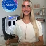
Self London has several resurfacing platforms, including UltraClear, HALO and erbium:YAG laser technology. Their value lies in allowing the treatment to be chosen according to the patient’s skin type, wrinkle depth, pigmentary risk and tolerance for recovery rather than forcing every concern onto one machine.
Someone with early crepiness and a low tolerance for downtime does not require the same treatment as a patient with established static wrinkles who is prepared for a more significant recovery. Periocular resurfacing requires appropriate eye protection and conservative judgement. Aggressive treatment may cause prolonged redness, pigmentary change, scarring or problems with lower-eyelid position.
Laser can produce a meaningful change in the surface and dermal quality of the skin. Orbital fat and skeletal anatomy remain where they were.
Botulinum toxin
Botulinum toxin has an established role in dynamic crow’s feet and can also be used strategically to influence muscular pull around the brow.[13] The dose and placement need to respect the patient’s smile and baseline brow position. Someone who relies on forehead contraction to lift a low brow may feel heavier if the frontalis is treated without recognising that compensation.
The best candidates have lines driven predominantly by movement and accept that normal expression should remain. Static crepiness may still be visible after the muscular component has been softened. A good result looks like a less sharply folded eye area when the patient smiles. It should not remove the smile from the eye.
Sofwave
Sofwave uses high-frequency ultrasound to create controlled thermal zones within the dermis, with the intention of stimulating collagen remodelling. Treatment across the forehead and brow may produce a modest improvement in early laxity and brow position. Device-specific clinical studies report improvement in facial and eyebrow laxity, although the evidence base is smaller and less mature than that supporting botulinum toxin or surgery.[14]
We consider Sofwave for patients with early brow descent or upper-face laxity who want a gradual, non-surgical improvement. The expected change is subtle and develops over several months.
It is not a treatment for lower-eyelid pigmentation, prominent orbital fat or true ptosis. Patients seeking a marked lift or removal of a significant fold of upper-eyelid skin are unlikely to be satisfied by an ultrasound treatment.
Tear-trough filler
Hyaluronic acid filler can improve a genuine structural hollow in a carefully selected patient. Selection is more important than the fact that the procedure can technically be performed. A relatively smooth lower eyelid, little tendency to swell and a clearly volume-related depression are more favourable than prominent bags, malar oedema, festoons or significant skin laxity.
Systematic reviews suggest that filler can improve tear-trough deformity, with commonly reported complications including bruising, oedema, contour irregularity and blue-grey discolouration.[9]
Direct injection beneath the eye is not always the most natural approach. Reduced mid-cheek support may be contributing to the lid-cheek transition. Existing filler may also be creating swelling or irregularity and should be identified before more is added.
Visual loss following facial filler injection is rare but potentially permanent. The risk cannot be reduced to zero, even with careful patient selection and technique.[18]
We would rather advise against tear-trough filler than create a persistently swollen lower eyelid in a patient whose anatomy was unsuitable from the outset.
Surgery
Significant upper-eyelid skin excess, prominent lower-eyelid fat or true ptosis may be better assessed by an oculoplastic or plastic surgeon. Upper blepharoplasty removes excess eyelid skin. Lower-eyelid surgery can address orbital fat and aspects of the lid-cheek junction. Ptosis surgery deals with the eyelid-elevation mechanism.
Surgery has its own risks and requires appropriate specialist assessment. It may still be the most proportionate treatment when the problem is clearly structural. A sequence of injectables and devices does not become equivalent to surgery because each step is described as non-invasive. Recommending an oculoplastic opinion can save a patient considerable time and money that would otherwise be spent pursuing a result those treatments were never capable of producing.
Three clinical patterns I see repeatedly
These are not individual case histories, but recurring patterns among patients who attend clinic for tired-looking eyes.
The patient treating a shadow as pigment
The patient has used brightening eye creams, retinoids and pigment serums, and may already have undergone peels or laser. The dark crescent remains.
When the face is examined in frontal daylight, much of the darkness disappears. Under overhead light, it becomes pronounced again. The dominant issue is the tear-trough contour and the way it casts a shadow rather than a large excess of melanin.
More pigment treatment is unlikely to create the change the patient wants. The options depend on the surrounding anatomy and may include no treatment, structural support in a carefully selected patient or acceptance that some normal shadow will remain.
The patient requesting filler beneath an eye that already swells
The patient notices a groove beneath the lower eyelid and assumes it needs to be filled. On examination, the stronger feature is the bag above it, combined with morning puffiness or malar oedema.
Placing water-binding hyaluronic acid into the region may soften part of the depression while making the whole lower eyelid appear heavier. Previous filler may already be contributing.
The most useful recommendation may be to avoid further filler, investigate whether old product remains and consider an oculoplastic opinion if orbital fat is the dominant concern.
The patient requesting polynucleotides for a structural problem
The patient has heard that polynucleotides are a natural or regenerative treatment for dark circles and expects them to remove a deep hollow or eye bag.
The skin may indeed be slightly crepey, but skin quality is not the feature driving the tired appearance. Improving hydration and surface texture will not substantially alter the contour.
Polynucleotides may still produce a modest improvement in the skin, but they are unlikely to satisfy the patient unless the structural limitation is understood in advance. Declining treatment may be more appropriate when the expected change does not match the concern.
Where treatment commonly goes wrong
Poor outcomes around the eyes are not always caused by poor products or devices. They often begin with a poorly defined target. A shadow is treated as pigment. A lower-eyelid bag is treated by filling the depression beneath it. A patient with dermatitis is given stronger active skincare. An upper-eyelid problem is treated beneath the eye. Thin skin is treated as though it were missing volume. The procedure may have been performed correctly and still be wrong for the face.
Another common problem is the accumulation of treatments. A patient has polynucleotides, then filler, then laser, then another skin booster within a short period. Neither patient nor practitioner can identify what helped, what was unnecessary or what caused the swelling or irritation.
Staging allows each response to be judged properly. Inflammation should settle before residual pigment is treated. Existing filler should be assessed before more is injected. Dynamic lines may be softened before deciding how much static skin change remains. Collagen-remodelling treatments need time to work.
Allowing each treatment sufficient time to work makes it easier to judge the result and avoid unnecessary escalation.
What can be done at home?
A sensible home routine can improve the skin around the eyes, provided its limitations are understood. Keep the eyelid barrier comfortable and avoid unnecessary irritation. A bland moisturiser is often more useful than a complex eye cream when the skin is dry or reactive. Introduce active ingredients individually rather than layering several together, and stop if itching, scaling or burning develops.
Retinoids may improve fine photoageing over time, but the lower eyelid is easily irritated. There is rarely a need to apply a strong retinoid directly to the lash line. Product placed around the orbital rim will migrate.
Treat allergy symptoms and try to reduce habitual rubbing. Simplifying eye make-up or identifying a contact allergen may reduce both inflammation and the mechanical stimulation of pigment.
Use broad-spectrum sunscreen and sunglasses. Where melasma or recurrent post-inflammatory hyperpigmentation is present, a tinted sunscreen containing iron oxides may offer additional visible-light protection.[5]
A cold compress can temporarily reduce morning puffiness. Regular sleep, moderate alcohol intake and avoiding an unusually salty meal may help fluid-related swelling in susceptible people.
These measures have much less influence over inherited pigment, orbital fat, a tear-trough ligament or brow position. Patients should not be encouraged to interpret every persistent feature around the eyes as proof that they are dehydrated, unhealthy or living incorrectly. Facial anatomy is not a lifestyle failure.
Take progress photographs in the same position, expression and lighting. A phone camera, ceiling spotlight or slight change in head position can create an apparent difference larger than the effect of many treatments.
Frequently asked questions
Why do my eyes look tired even after eight hours of sleep?
Sleep can improve temporary redness, swelling and dullness. It cannot alter inherited pigmentation, translucent skin, a tear-trough ligament, orbital fat or brow position. Where the appearance persists despite adequate rest, anatomy and skin biology are usually more important than the number of hours slept.
How can I tell whether my dark circles are caused by pigment or shadow?
Pigment tends to remain visible as the lighting changes. A contour shadow becomes stronger beneath overhead light and often softens when the face is illuminated directly from the front. Blue or purple colour may reflect visible vessels or underlying muscle. These clues are useful, but mixed cases are common and are best assessed in person.
Why do my dark circles look worse in photographs?
A camera converts a three-dimensional structure into a flat image. Phone processing may increase contrast, while overhead lighting creates a deeper shadow beneath the orbital rim, lower-eyelid bag or tear trough. The photograph may be recording the lighting rather than revealing a sudden deterioration in the skin.
Can dark circles be removed completely?
Some components can be improved substantially when there is a clear and treatable cause. Complete removal is less realistic when inherited pigment, translucent skin and structural shadowing coexist. The eye is a three-dimensional structure and will continue to cast normal shadows in ordinary light.
Are polynucleotides worth having beneath the eyes?
They can be worthwhile when the main concern is fine, crepey or fragile-looking skin and the patient is comfortable with a gradual, modest change. They are much less likely to satisfy someone whose dominant concern is a deep hollow, prominent eye bag or significant upper-eyelid heaviness.
Will tear-trough filler remove dark circles?
It may soften darkness caused mainly by a genuine structural hollow in suitable anatomy. It does not remove melanin, visible vessels or crepey skin. Patients with orbital fat bags, malar oedema, festoons or a tendency to swell may find that filler makes the lower eyelid look heavier rather than more rested.
What is the best laser for crepey under-eye skin?
There is no single best device. The choice depends on skin type, wrinkle depth, pigmentary risk, previous treatment and the amount of recovery the patient can accept. The wavelength matters, but the parameters and the judgement of the practitioner matter just as much.
Can Sofwave lift hooded eyes?
Sofwave may produce a modest improvement when early brow descent or upper-face laxity contributes to heaviness. It cannot remove a substantial fold of upper-eyelid skin or correct true ptosis. Patients seeking a marked anatomical change may require an oculoplastic assessment.
Are eye bags caused by poor sleep?
Poor sleep can increase temporary puffiness, particularly in the morning. Persistent bags more commonly reflect inherited or age-related prominence of orbital fat. A patient can sleep well and still have eye bags because the underlying problem is structural.
When should a change around the eyes be assessed medically?
A sudden eyelid droop, new one-sided swelling, pronounced redness, eye pain, double vision or any change in sight requires medical assessment. These are not routine cosmetic signs of tiredness or ageing and should not be managed with an aesthetic procedure.[17]
Assessment and treatment at Self London
A consultation for tired-looking eyes at Self London on Harley Street examines the whole periocular area rather than assuming that the answer lies in a particular injection or device.
The assessment includes pigmentation, eyelid inflammation, vascular visibility, skin texture, tear-trough and cheek anatomy, fluid retention, lower-eyelid fat, brow position, muscular movement and previous treatment. These findings are considered together because an intervention aimed at one part of the problem may have little effect on the feature the patient is actually noticing.
A patient with post-inflammatory pigmentation may need control of eyelid dermatitis and a carefully planned pigment regimen. Someone with crepey lower-eyelid skin may be suited to polynucleotides or laser resurfacing. Dynamic crow’s feet may respond well to conservative botulinum toxin, while early brow descent may be appropriate for Sofwave. A patient with prominent orbital fat or significant upper-eyelid skin excess may receive more useful advice from an oculoplastic surgeon than from another non-surgical procedure.
Self London is a consultant dermatologist-led, CQC-regulated medical and aesthetic clinic with access to several laser and energy-based technologies. The advantage of having more than one platform is not the ability to offer more procedures. It is the ability to select a treatment according to the skin, anatomy and tolerance for recovery rather than fitting every concern to the same machine.
The patients who tend to do best have a clearly identified target and realistic expectations. Fine, crepey skin can often be improved. Dynamic crow’s feet can be softened. Selected pigmentation and vascular colour may respond to targeted treatment. Early brow laxity may improve modestly, and some structural hollows can be corrected in carefully chosen patients.
The result should make the eye area look fresher without leaving it swollen, immobile or obviously treated. Occasionally, the likely improvement is too limited to justify the cost, recovery or risk. Patients should be told that before, rather than after, a procedure.
Persistent tired-looking eyes may be caused by colour, contour, fluid, skin quality or the position of the tissues around them. Once those components have been separated, it becomes much easier to decide whether skincare, laser, polynucleotides, botulinum toxin, Sofwave, filler or surgery has a credible role.
Patients concerned about dark circles, under-eye crepiness, eye bags or upper-eyelid heaviness can book a consultation at Self London for a full periocular assessment.
References
- Sundelin T, Lekander M, Kecklund G, van Someren EJW, Olsson A, Axelsson J. Cues of fatigue: effects of sleep deprivation on facial appearance. Sleep. 2013;36(9):1355–1360. PMID: 23997369.
- Michelle L, Pouldar Foulad D, Ekelem C, Saedi N, Atanaskova Mesinkovska N. Treatments of periorbital hyperpigmentation: a systematic review. Dermatologic Surgery. 2021;47(1):70–74. PMID: 32740208.
- Sarkar R, Ranjan R, Garg S, Garg VK, Sonthalia S, Bansal S. Periorbital hyperpigmentation: a comprehensive review. Journal of Clinical and Aesthetic Dermatology. 2016;9(1):49–55. PMID: 26962392.
- Borzova E, Snarskaya E, Bratkovskaya A, et al. Eyelid dermatitis in patch-tested adult patients: a systematic review with a meta-analysis. Scientific Reports. 2024;14:18791. PMID: 39138344.
- Lyons AB, Trullas C, Kohli I, Hamzavi IH, Lim HW. Photoprotection beyond ultraviolet radiation: a review of tinted sunscreens. Journal of the American Academy of Dermatology. 2021;84(5):1393–1397. PMID: 32335182.
- Wong CH, Hsieh MKH, Mendelson B. The tear trough ligament: anatomical basis for the tear trough deformity. Plastic and Reconstructive Surgery. 2012;129(6):1392–1402. PMID: 22634656.
- Liew S, Doreian S, Kunathathorn W, et al. Lower Eyelid Dark Circles (Tear Trough and Lid-Cheek Junction): A Stepwise Assessment Framework. Aesthetic Surgery Journal. 2024;44(7):NP476–NP485. PMID: 38489829.
- Mustak H, Fiaschetti D, Goldberg RA. Filling the periorbital hollows with hyaluronic acid gel: long-term review of outcomes and complications. Journal of Cosmetic Dermatology. 2018;17(4):611–616. PMID: 29130598.
- Trinh LN, Gupta A. Dermal fillers for tear trough rejuvenation: a systematic review. Facial Plastic Surgery. 2022;38(3):228–239. PMID: 34192769.
- Lee YJ, Kim HT, Lee YJ, et al. Comparison of the effects of polynucleotide and hyaluronic acid fillers on periocular rejuvenation: a randomised, double-blind, split-face trial. Journal of Dermatological Treatment. 2022;33(1):254–260. PMID: 32248707.
- Lampridou S, Bassett S, Cavallini M, Christopoulos G. The Effectiveness of Polynucleotides in Esthetic Medicine: A Systematic Review. Journal of Cosmetic Dermatology. 2025;24(2):e16721. doi:10.1111/jocd.16721. PMID: 39645667.
- Karsai S, Czarnecka A, Jünger M, Raulin C. Ablative fractional lasers (CO₂ and Er:YAG): a randomized controlled double-blind split-face trial of the treatment of peri-orbital rhytides. Lasers in Surgery and Medicine. 2010;42(2):160–167. doi:10.1002/lsm.20879. PMID: 20166156.
- Camargo CP, et al. Botulinum toxin type A for facial wrinkles. Cochrane Database of Systematic Reviews. 2021. PMID: 34224576.
- Gold MH, Biron J. Efficacy and safety of high-intensity, high-frequency, non-focused ultrasound parallel beams for facial skin laxity. Journal of Cosmetic Dermatology. 2024;23(1):117–123. PMID: 38031530.
- Park M, Akuffo-Addo E, Mar K, Zhu CK, Mukovozov I. Treatments for periorbital hyperpigmentation in Fitzpatrick skin types IV–VI: a systematic review. Archives of Dermatological Research. 2024;316(8):552. PMID: 39172264.
- Beer J, et al. What’s new with under-eye treatment: a multispecialty systematic review of recent under-eye treatments. Dermatologic Surgery. 2026. PMID: 41615388.
- NHS. Eyelid problems. Guidance on symptoms requiring urgent medical assessment.
- Kapoor KM, Kapoor P, Heydenrych I, Bertossi D. Vision loss associated with hyaluronic acid fillers: a systematic review of literature. Aesthetic Plastic Surgery. 2020;44(3):929–944. PMID: 31822960.